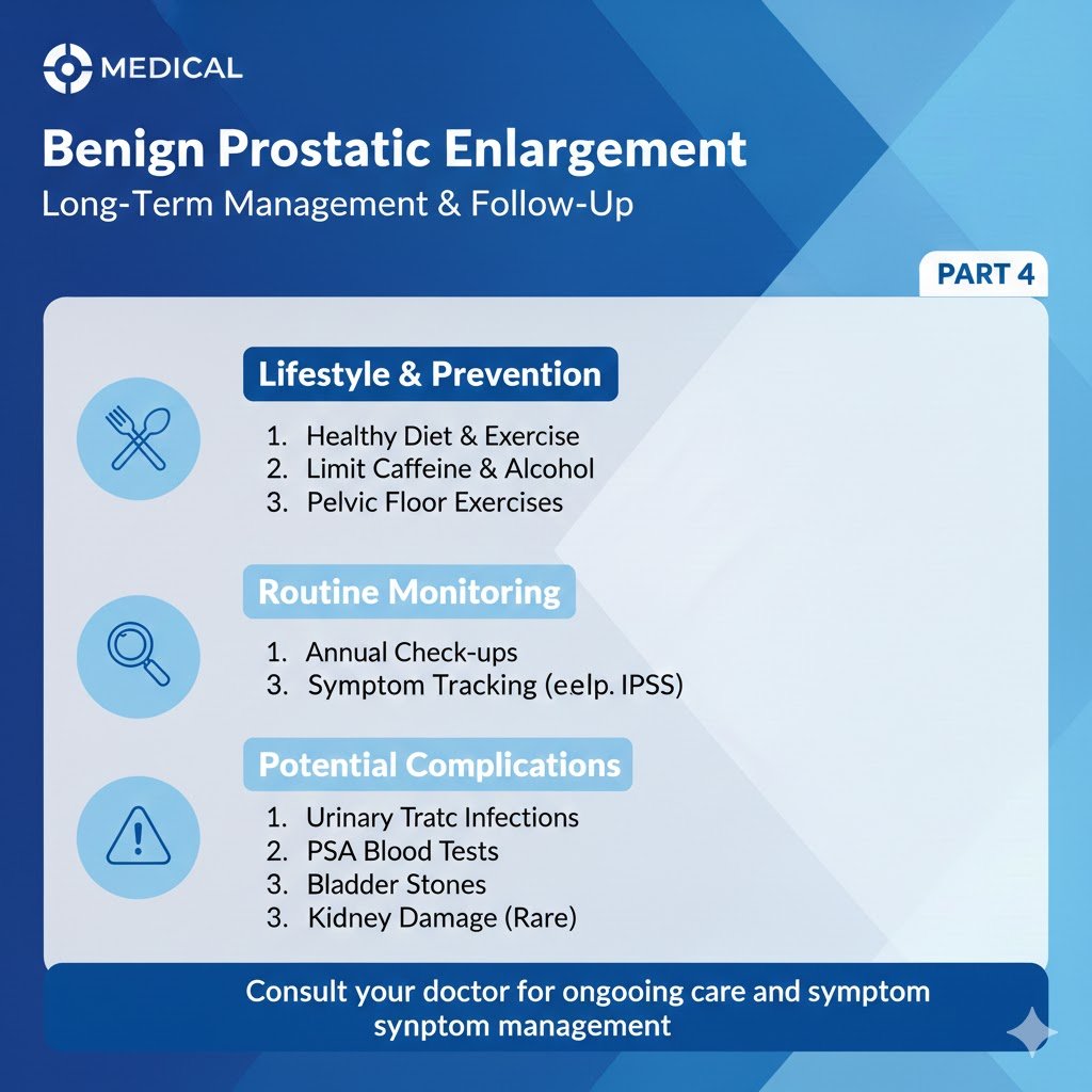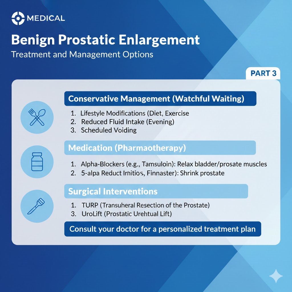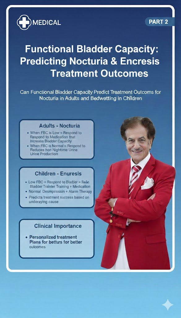
هل يمكن للسعة الوظيفية للمثانة أن تتنبأ بنتائج علاج التبول الليلي لدى الرجال والنساء والسلس البولي الليلي لدى الاطفال؟ ج2
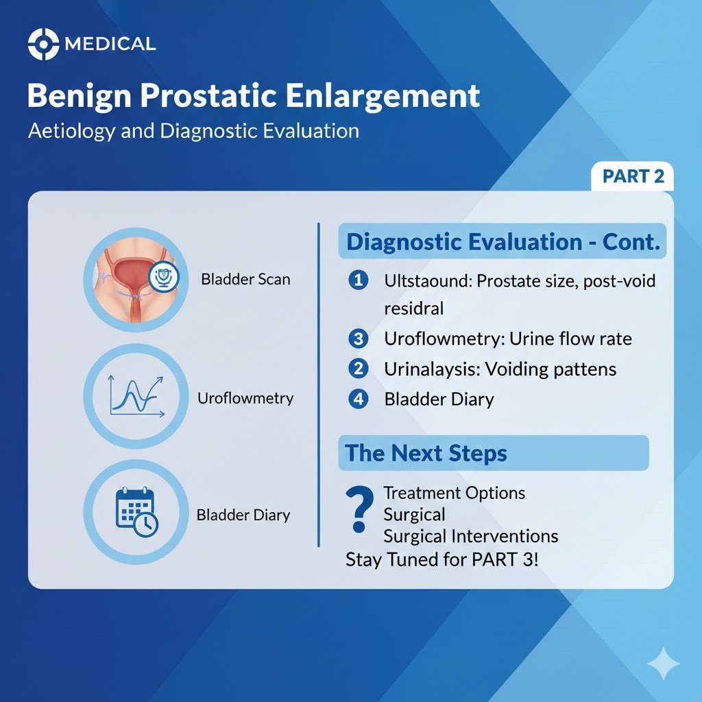
هل يمكن للسعة الوظيفية للمثانة أن تتنبأ بنتائج علاج التبول الليلي لدى الرجال والنساء والسلس البولي الليلي لدى الاطفال؟ الجزء الثاني البروفيسور الدكتور سمير أحمد السامرائي أما إصابة الأطفال بالبيلة الليلية فإنه تم تعريفها بأنها سلس بولي ليلي (NE) مع عدم السيطرة الانتظامية على التبول وحدوثه ويكون غير إرادي أثناء النوم وأثناء الليل وخاصة لدى الأطفال الذين تتراوح أعمارهم ما بين 5 سنوات و18 سنة. يقسم السلس البولي الليلي إلى نوعين أولهما سلس بولي ليلي ذو العرض الواحد (MNE) وثانيهما سلس بولي ليلي الذي يظهر مع أعراض أخرى في المسالك البولية السفلى (NMNE)، أما الأطفال المصابون بالنوع الثاني فإنهم مصابون أيضا بإضطرابات وظيفية في المثانة، وقد يؤدي هذا السلس البولي الليلي لهؤلاء الأطفال إلى مشاكل نفسية ومشاكل في حياتهم الإجتماعية اليومية. يعد سلس البول الليلي بأنه مرض متعدد العوامل. الأسباب الرئيسية فإنها تكون من جراء التبول الليلي الزائد (Nocturnal Polyuria) بسبب الإصابة جينيا أو مكتسبا بإنخفاض إنتاج الهرمون المانع للإدرار (ADH) وهذا الهرمون هو المسؤول على توازن السوائل في الجسم، أو الإصابة بالمثانة ذو السعة الصغيرة (Low Functional Bladder Capacity) وكذلك الإصابة بإضطراب الإستثارة أثناء النوم أو إصابتهم وراثيا من أحد الوالدين نسبة 40% أو كلاهما بنسبة 70%. تشخيص وتقييم سعة المثانة الوظيفية لدى هؤلاء الأطفال المصابون بالبيلة الليلية يتم من خلال معرفة وتثبيت المعلومات من الطفل وعائلته وخاصة عن تبوله أثناء النهار وأثناء الليل مع قياس سعة المثانة بواسطة الموجات فوق الصوتية وديناميكيتها بواسطة جهاز الذبذبات التفريغية البولية (Uroflowmetry) وكذلك قياس سمك جدار المثانة وقياس المتبقي في المثانة بعد التبول. هذه الوسائل التشخيصية تعتبر أمرا أساسيا في وضع الإستراتيجية العلاجية لهؤلاء الأطفال ومراقبة إستجابتهم لهذا العلاج. رغم الدراسات العديدة حول السلس البولي الليلي، فإنه لايزال هنالك تباين كبير في الطريقة التشخيصية والتقييمية وكذلك الإستراتيجيات العلاجية لهذا المرض. تشخيصيا توصي جمعية السلس البولي لدى الأطفال لتخمين وتقييم وتشخيص صغر سعة المثانة الوظيفية (FBC) مع استخدام مخططات التردد إلى الحمام للتبول ليلا ونهارا وقياس كمية البول خلال 48 ساعة (48-h F/V) حيث أنه من خلال هذه الطريقة التشخيصية نستطيع أن نقيم قدرة وسعة المثانة الوظيفية وكذلك نستطيع معرفة كمية البول القصوى (MVV) الذي يمكن الحصول عليه بإستخدام هذه المخططات (48-h F/V charts) لمعرفة القدرة الوظيفية للمثانة الحقيقية. ومن الوسائل التشخيصية المهمة كذلك هو تشخيص المتبقي من البول في المثانة بعد تفريغها بواسطة فحص المثانة بالموجات فوق الصوتية وقياس البول المتبقي، إضافة إلى ذلك تقاس ديناميكية تدفق البول (UFM) من خلال الإحليل [1] وكذلك قد نلجأ إذا لم يتم تشخيص السبب الرئيسي لهذا المرض إلى فحص تفريغ المثانة بواسطة التصوير بالأشعة السينية (Micturating Cystography)، وهذه تعتبر كوسائل تشخيصية بديلة لتقييم القدرة الوظيفية للمثانة. أظهرت العديد من الدراسات أن سعة المثانة الوظيفية تقل إلى 50% لدى هؤلاء الأطفال الذين يعانون من التبول الليلي. ووفقًا لدراسة Kim، كان 46.5٪ من جميع هؤلاء الأطفال لديهم سعة مثانية مخفضة بالنسبة لأعمارهم، وإرتفع تشخيص سعة مثانية صغيرة لدى هؤلاء الأطفال الذين يشكون من التبول اللاإرداي في الفراش يوميا ولدى الأطفال الذين يتبولون في الفراش أثناء الليل لمرات عديدة. في دراسةLiu et al ، كان 33.9٪ من هؤلاء الأطفال الذين يعانون من التبول الليلي الأحادي مصابون بصغر سعة المثانة. وأفادت دراسة Acosta et al بأن 85٪ من هؤلاء الاطفال المصابون بالتبول اللاإرادي ليلا شخص إصابتهم بسعة مثانية صغيرة وأقل من 70٪ من السعة المتوقعة للمثانة في أعمارهم، وربما يعود ذلك إلى أن الحد الأدنى للسعة الوظيفية الطبيعية للمثانة وضع في 70%. وذكر Kang وآخرون بأن السعة الوظيفية تكون 68٪-70٪ (وفقًا لطرق القياس) بأن هؤلاء الأطفال لديهم سعة مثانية صغيرة بالنسبة لأعمارهم، بغض النظر عن مجموعة التبول الليلي .[2] أظهرت عدة دراسات بأن علاج هؤلاء الأطفال بالهرمون المضاد للإدرار (Desmopressin) قد أثرعلى السعة الوظيفية للمثانة. حيث أفاد الباحث كيم عن وجود علاقة ملحوظة بين شدة التبول الليلي الغير إرادي ودرجة انخفاض السعة الوظيفية للمثانة .[3] كما وأفاد الباحث يونج وآخرون بأن مجموعة هؤلاء الأطفال ذو السعة الوظيفية الصغيرة يتعرضون لإنتكاسات في الاستجابة أثناء العلاج بالهرمون المضاد للإدرار (الديسموبريسين) [4]. وكما ذكر إيلر [5] ورشتون [6] أنه إذا تجاوزت السعة الوظيفية للمثانة 70% من السعة المتوقعة والمعدلة لعمر الطفل، فمن المتوقع أن يكون هناك استجابة جيدة للعلاج بواسطة الهرمون المضاد للإدرار (الديسموبريسين)، ومع ذلك فقد أكدت دراسة إكلينيكية حديثة بإرتفاع السعة الوظيفية للمثانة بنسبة 30% أو أكثر ورغم توقف العلاج لمدة 6 أشهر. وفي الأخير تشير المبادئ التوجيهية العملية لعلاج التبول الليلي اللاإرادي عند هؤلاء الأطفال إلى أن السعة الوظيفية المنخفضة للمثانة مقارنة بالعمر كان لها علاقة بالإستجابة المنخفظة لعلاج الهرمون المضاد للإدرار (الديمسوبريسين). ولكن لم يجد Chang و Yang أي علاقة ملحوظة بين السعة الوظيفية المنخفضة للمثانة والاستجابة للعلاج هذا، بل أكدوا بدلاً من ذلك بأن المتبقي من البول بعد تفريغ المثانة و إرتفاع نسبته وفي نفس الوقت إرتفاع نسبة الإدرار بكمية كبيرة أثناء الليل كانت عوامل تنبؤية مهمة للإستجابة إلى علاج هذا المرض. وفي دراسة للباحث Liu وآخرين، دلت على أن السعة الوظيفية للمثانة لم تكن عاملًا تنبؤيًا للاستجابة على علاج الهرمون المضاد للإدرار [7]. في الخلاصة، يعدُّ الاضطراب اللاإرادي للتبول الليلي لدى هؤلاء الأطفال مرضًا معقدًا له عوامله المذكورة آنفا. ولهذا فإن إستخدام الوسائل التشخيصية العديدة لمعرفة سعة المثانة الوظيفية وعلاقتها بالسلس الليلي اللاإرادي عند هؤلاء الأطفال تعد هدفا تشخيصيا أوليا لتحديد إستراتيجية العلاج. ويعد الرسم البياني F/V لمدة 48 ساعة والاختبار الوظيفي للمثانة مع قياس المتبقي بالمثانة بعد تفريغها وسائل تشخيصية موثوقة لقياس سعة المثانة الوظيفية لدى هؤلاء الاطفال الذين يعانون من السلس البولي الليلي وخاصة الذين يعانون من السلس البولي الليلي الشديد فقد أضهرت بأن السعة الوظيفية للمثانة تعتبر علامة تشخيصية مهمة لعلاج التبول الليلي اللاإرادي عند هؤلاء الأطفال والذي يجب أن يكون ليس أحاديا بالهرمون المضاد للإدرار وإنما مركبا يحتوي على علاج المثانة ذو الفعالية الفائقة وذو السعة الوظيفية الصغيرة لكي تكون نسبة نجاحه عالية كما أثبتتها الدراسات المذكورة آنفا.Top of Form REFERENCES: [1]: Maternik M, Chudzik I, Krzeminska K et al: Evaluation of bladder capacity in children with lower urinary tract symptoms: comparison of 48-hour frequency/volume charts and uroflowmetry measurements. J Pediatr Urol 2016; 12: 214.e1. [2]: Kang BJ, Chung JM and Lee SD: Evaluation of functional bladder capacity in children with nocturnal enuresis according to type and treatment outcome. Res Rep Urol 2020; 12: 383. [3]: Kim JM: Diagnostic value of functional bladder capacity, urine osmolality, and daytime storage symptoms for severity of nocturnal enuresis. Korean J Urol 2012; 53: 114.