أفضل مركز مسالك بولية دبي

Overactive Urinary Bladder

Overactive Urinary Bladder

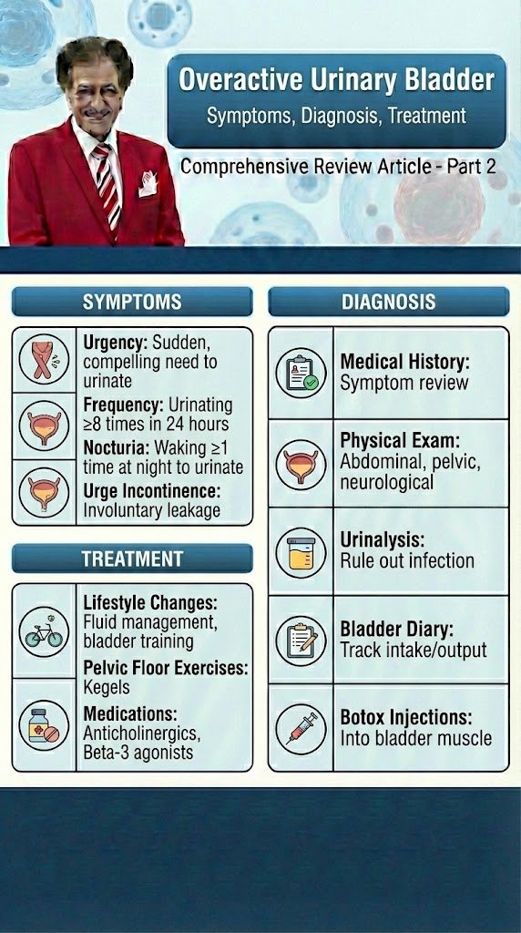
Overactive Urinary Bladder Diagnostic and Evaluation Comprehensive Review Article Part 2 Prof. Dr. Semir. A. Salim. Al Samarrai Taking a thorough clinical history is fundamental to the process of clinical evaluation. Despite the lack of high-level evidence to support taking a history, there is universal agreement that it should be the first step in the assessment of anyone with lower urinary tract symptoms (LUTS). The history should include a full evaluation of LUTS, as well as sexual, gastrointestinal and neurological symptoms. Details of urgency episodes, the type, timing and severity of urinary incontinence (UI), and some attempt to quantify symptoms should also be made. The history should help to categorise LUTS as storage, voiding and post-micturition symptoms, and classify UI as stress urinary incontinence (SUI), urge urinary incontinence (UUI), mixed urinary incontinence (MUI) or overflow incontinence; the latter being defined as “the complaint of UI in the symptomatic presence of an excessively (over-) full bladder (no cause identified)” [1]. The history should also identify patients who need referral to an appropriate clinic/specialist. These may include patients with associated pain, haematuria, history of recurrent urinary tract infection (UTI), pelvic surgery or radiotherapy, constant leakage suggesting a fistula, new-onset enuresis or suspected neurological disease. A neurological, obstetric and gynaecological history may help to understand the underlying cause and identify factors that may affect treatment decisions. Guidance on history-taking and diagnosis in relation to UTIs, neuro-urological conditions and chronic pelvic pain (CPP) can be found in the relevant EAU Guidelines [2,3,4]. Patients should also be asked about other comorbidity as well as smoking status, previous surgical procedures and current medications, as these may affect LUTS. There is little evidence from clinical trials that carrying out a clinical examination improves outcomes, but widespread consensus suggests that clinical examination remains an essential part of assessment of patients with LUTS. Examination should include abdominal examination, to detect an enlarged urinary bladder or other abdominal mass, and digital examination of the vagina and/or rectum. Pelvic examination in women includes assessment of oestrogen status, pelvic floor muscle (PFM) function and careful assessment of any associated pelvic organ prolapse (POP). A cough stress test is necessary to look for stress urinary incontinence (SUI). Among women with genital prolapse, the cough test was found to show good agreement with urodynamic studies (UDS) in the detection of SUI. Urethral mobility can be assessed. Pelvic floor muscle contraction strength can also be assessed digitally. A focused neuro-urological examination should also be routinely undertaken. Patient questionnaires include symptom scores, symptom questionnaires/scales/indices, patient-reported outcome measures (PROMs) and health-related quality of life (QoL) measures. Questionnaires should have been validated for the language in which they are being used, and, if used for outcome evaluation, should have been shown to be sensitive to change. The US Food and Drug Administration (FDA) published guidance for industry on PROM instruments (questionnaires) in 2009 [5]. Patient bladder diaries include measurement of the frequency and severity of LUTS and is an important step in the evaluation and management of LUT dysfunction. Bladder diaries are a semi-objective method of quantifying symptoms, such as frequency of urinary incontinence (UI) events, number of nocturia episodes, etc. Fluid intake and voided volume measurement can be used to support diagnoses and management planning, for example in overactive bladder (OAB), and for identifying 24-hour or nocturnal polyuria. The optimum number of days required for bladder diaries appears to be based on a balance between accuracy and compliance [6,7]. Diary durations between three and seven days are routinely reported in the literature. The urinalysis and urinary tract infection investigations are a very important steps in the evaluation and therapy of UTI. Reagent strip (dipstick) urinalysis may indicate proteinuria, haematuria or glycosuria, or suggest UTI requiring further assessment. Urine dipstick testing is a useful adjunct to clinical evaluation in patients in whom urinary symptoms are suspected to be due to UTI. Urinalysis negative for nitrite and leukocyte esterase may exclude bacteriuria in women with LUTS [8], and should be included, with urine culture when necessary, in the evaluation of all patients with LUTS. Urinary incontinence or worsening of LUTS may occur during UTI [9] and existing UI may worsen [10]. The rate and severity of UI were unchanged after eradication of asymptomatic bacteriuria in nursing home residents [11]. The post-void residual volume measurement is also important step in the evaluation and management of OAB and obstruction LUT-Disorders. Post-void residual (PVR) volume is the amount of urine that remains in the bladder after voiding. It is a measure of voiding efficiency, and results from a number of contributing factors. The detection of significant PVR volume is important because it may worsen symptoms and, more rarely, may be associated with UTI, upper urinary tract (UUT) dilatation and renal insufficiency. Both BOO and/or detrusor underactivity (DU) can potentially contribute to the development of significant PVR volume. Post-void residual volume can be measured by catheterisation or ultrasound (US). Most studies investigating PVR volume have assessed mixed populations including those with neurogenic UI. In general, the data on PVR volume can be applied with caution to women with non-neurogenic LUTS. The results of studies investigating the best method of measuring PVR volume [11-16] have led to the consensus that US measurement of PVR volume is preferable to catheterisation due to its favourable risk–benefit profile. In peri- and postmenopausal women without significant LUTS or pelvic organ symptoms, 95% had a PVR volume < 100 mL [17]. In women with UUI, PVR volume > 100 mL was found in only 10% of cases [18]. Other research has found that a high PVR volume is associated with pelvic organ prolapse (POP), voiding symptoms and an absence of SUI [17, 19–21]. In women with SUI, the mean PVR volume was 39 mL measured by catheterisation and 63 mL measured by US, with 16% of women having PVR volume > 100 mL [22]. Some authors have suggested that it is reasonable to consider a PVR volume > 100 mL to be significant, although many women may remain

Overactive Urinary Bladder قراءة المزيد »

Overactive Urinary Bladder

Overactive Urinary Bladder

Overactive Urinary Bladder Epidemiology, aetiology, pathophysiology Comprehensive Review Article Part 1 Prof. Dr. Semir. A. Salim. Al Samarrai Overactive bladder is defined by the International Continence Society (ICS) as “urinary urgency, usually accompanied by frequency and nocturia, with or without (Urge Urinary Incontinence (UUI), in the absence of Urinary Tract Infection (UTI) or other obvious pathology” [1]. Overactive bladder is a chronic condition and can have debilitating effects on QoL. The hallmark urodynamic feature is Detrusor Overactivity (DO), although this may not be demonstrated in a large proportion of Overactive Bladder (OAB) patients, which may partly be due to failure to reproduce symptoms during urodynamic assessment. The EPidemiology of InContinence (EPIC) study was one of the largest population-based surveys of the prevalence of LUTS and OAB [2]. It was a cross-sectional telephone survey of adults aged > 18 years conducted in five countries, including Canada, Germany, Italy, Sweden and the UK. The study included > 19,000 participants and demonstrated an overall prevalence of OAB symptoms of 11.8% (10.8% in men and 12.8% in women). Other studies have reported prevalences of up to 30 to 40%, with rates generally increasing with age [3]. Various theories have been proposed to explain the pathophysiology of OAB, mainly relating to imbalances in inhibitory and excitatory neural pathways to the bladder and the urethra or sensitivity of bladder muscle receptors. However, no definite identifiable causes have been established. Overactive bladder is generally classified into wet and dry, based on the presence or absence of associated Urinary Incontinence (UI). Evaluation of symptoms of OAB follows the general pathway of evaluation of women with LUTS. The Diaries are particularly helpful in establishing and quantifying symptoms of frequency, urgency and UI, and may be valuable in assessing change over time or response to treatment. Several observational studies have demonstrated a close correlation between data obtained from bladder diaries and standard symptom evaluation [4–7]. The optimum number of days required for bladder diaries appears to be based on a balance between accuracy and compliance. Diary duration of three to seven days is routinely used in the literature. The Urodynamics is essential in establishing the presence of DO, but its absence does not preclude diagnosis of OAB, which is based on symptoms alone. A Cochrane review of seven RCTs showed that use of urodynamic tests increased the likelihood of prescribing drugs or avoiding surgery. However, there was no evidence that this influence on decision-making altered the clinical outcome of treatment [8]. A sub-analysis of an RCT comparing fesoterodine to placebo [9] showed that the urodynamic diagnosis of DO had no predictive value for treatment response. A single report (SR) and meta-analysis indicated that the urinary tract nerve growth factor (Urinary NGF) and Brain-divided neurotrophic factor or abneurin are members of the neurotrophic family of growth factors were increased in female OAB patients as urinary biomarker compared to healthy controls, whereas no difference was found for the prostaglandins E2 (PGE2) level normalized to the concentration of the urinary creatine was elevated and higher in the BPH/OAB than in the BPH/non-groups [10]. The current data is inadequate to assess any other potential biomarkers, such as urinary malondialdehyde (UMDA), ATP, and cytokines, in the management of OAB in female patients. Further studies are needed to establish their potential as diagnostic and management tools in OAB women. The conservative management of the overactive bladder has long been recommended as first in clinical practice, because they usually carry the lowest risk of harm. While this remains true for non-pharmacological conservative treatments [e.g., pelvic floor muscle training (PFMT)], increasing concerns regarding the adverse events of some pharmacological treatments used to treat LUTS (e.g., anticholinergic drugs), particularly regarding cognitive function, have emerged and patients should be fully counselled regarding this potential risk. It is possible that improvement of associated disease may reduce the severity of the lower urinary tract symptoms (LUTS), especially in elderly patients, which are associated with multiple comorbid conditions including: • cardiac failure; • chronic renal failure; • diabetes; • chronic obstructive pulmonary disease; • neurological disease; • general cognitive impairment; • sleep disturbances, e.g., sleep apnoea; • depression; • metabolic syndrome. The Lifestyle factors that may be associated with UI include obesity, smoking, level of physical activity, regulation of bowel habit and fluid intake. Modification of these factors may improve symptoms of OAB. The caffeine intake in many drinks contain caffeine are particularly coffee, tea and cola. Conflicting epidemiological evidence of urinary symptoms being aggravated by caffeine intake has focused on whether caffeine reduction improves LUTS [11, 12]. A scoping review of fourteen interventional and twelve observational studies reported that reduction in caffeine intake may reduce symptoms of urgency, but the certainty of evidence was low, with significant heterogeneity in study populations [13]. The fluid intake modification are particularly restriction, and is a strategy commonly used by people with OAB to relieve symptoms. Any advice on fluid intake given by HCPs should be based on 24-hour fluid intake and urine output measurements as retrieved from the bladder diary. From a general health point of view, it should be advised that fluid intake should be sufficient to avoid thirst and that an abnormally low or high 24-hour urine output should be investigated. The few RCTs that have been published provide inconsistent evidence [14-16]. In most studies, the instructions for fluid intake were individualised and it was difficult to assess participant adherence. All available studies were in women. An RCT showed that a reduction in fluid intake by 25% improved symptoms in patients with OAB but not UI [16]. Personalised fluid advice compared to generic advice made no difference to continence outcomes in people receiving anticholinergics for OAB, according to an RCT comparing drug therapy alone to drug therapy with behavioural advice [17]. Patients should be warned of the potential consequences of fluid restriction such as worsening of constipation or development of UTI. The obesity and overweight have been identified as a risk factors for LUTS in many epidemiological studies [18, 19]. There is

Overactive Urinary Bladder قراءة المزيد »

Prostate Cancer Salvage Therapy Comprehensive Review Article Part 7

Prostate Cancer Salvage Therapy Comprehensive Review Article Part 7

Prostate Cancer Salvage Therapy Comprehensive Review Article Part 7 Prof. Dr. Semir. A. Salim. Al Samarrai Salvage radiotherapy combined with androgen deprivation therapy (cTxcN0, without PET/CT): Data from RTOG 9601 suggest both CSS and OS benefit when adding 2 years of bicalutamide (150 mg o.d.) to SRT [1]. However, SRT combined with either goserelin or placebo showed similar DSS and OS rates [2]. Table 1 provides an overview of these two RCTs. Table 1. Randomised controlled trials comparing salvage radiotherapy combined with androgen deprivation therapy vs. salvage radiotherapy alone Target volume, dose, toxicity There have been various attempts to define common outlines for ‘clinical target volumes‘ of PCa [3–6] and for organs at risk of normal tissue complications [7]. A benefit in biochemical PFS but not metastasis-free survival has been reported in patients receiving whole pelvis SRT (± ADT) but the advantages must be weighed against possible side effects [8]. Two RCT’s were recently published (Table 2). Intensity-modulated radiation therapy plus IGRT was used in 57% of the patients in the SAKK-trial [9] and in all patients of the Chinese trial [10]. No patient had a PSMA PET/CT before randomisation. Table 2. Randomized trials investigating dose escalation for SRT without ADT and without PET-CT Salvage RT is associated with toxicity. In one report on 464 SRT patients receiving median 66.6 (max. 72) Gy, acute grade 2 toxicity was recorded in 4.7% for both the GI and GU tract. Two men had late grade 3 reactions of the GI tract, but overall, severe GU tract toxicity was not observed. Late grade 2 complications occurred in 4.7% (GI tract) and 4.1% (GU tract), respectively, and 4.5% of the patients developed moderate urethral stricture [11]. Salvage RT with or without ADT (cTx CN0/1) with PET/CT In a prospective multi-centre study of 323 patients with BCR, PSMA PET/CT changed the management intent in 62% of patients as compared to conventional staging. This was due to a significant reduction in the number of men in whom the site of disease recurrence was unknown (77% vs. 19%, p < 0.001) and a significant increase in the number of men with metastatic disease (11% vs. 57%) [12]. Metastasis-directed therapy for rcN+ (with PET/CT) Radiolabelled PSMA PET/CT is increasingly used as a diagnostic tool to assess metastatic disease burden in patients with BCR following prior definitive therapy. A review including 30 studies and 4,476 patients showed overall estimates of positivity in a restaging setting of 38% in pelvic LNs and 13% in extra-pelvic LN metastases [13]. The percentage positivity of PSMA PET/CT was proven to increase with higher PSA values, from 33% (95% CI: 16–51) for a PSA of < 0.2 ng/mL, to 45% (39–52), 59% (50–68), 75% (66–84), and 95% (92–97) for PSA subgroup values of 0.2–0.49, 0.5–0.99, 1.00–1.99, and > 2.00 ng/mL, respectively [13]. Salvage lymph node dissection The surgical management of (recurrent) nodal metastases in the pelvis has been the topic of several retrospective analyses [14-16] and a systematic review [17]. The reported 5-year BCR-free survival rates ranged from 6% to 31%. Five-year OS was approximately 84% [17]. Biochemical recurrence rates were found to be dependent on PSA at surgery and location and number of positive nodes [18]. Addition of RT to the lymphatic template after salvage LN dissection may improve the BCR rate [19]. Management of PSA failures after radiation therapy Therapeutic options in these patients are ADT or salvage local procedures. A systematic review and metaanalysis included studies comparing the efficacy and toxicity of salvage RP, salvage HIFU, salvage cryotherapy, SBRT, salvage LDR brachytherapy, and salvage HDR brachytherapy in the management of locally recurrent PCa after primary radical EBRT [20]. The outcomes were BCR-free survival at 2 and 5 years. Salvage radical prostatectomy Salvage RP after RT is associated with a higher likelihood of adverse events (AEs) compared to primary surger because of the risk of fibrosis and poor wound healing due to radiation [21]. Oncological outcomes In a systematic review of the literature, Chade, et al., showed that SRP provided 5- and 10-year BCR-free survival estimates ranging from 47–82% and from 28–53%, respectively. The 10-year CSS and OS rates ranged from 70–83% and from 54–89%, respectively. Pathological T stage > T3b (OR: 2.348) and GS (up to OR 7.183 for GS > 8) were independent predictors for BCR (see Table 3). Table 3. Oncological results of selected salvage radical prostatectomy case series Morbidity Compared to primary open RP, SRP is associated with a higher risk of later anastomotic stricture (47 vs. 5.8%), urinary retention (25.3% vs. 3.5%), urinary fistula (4.1% vs. 0.06%), abscess (3.2% vs. 0.7%) and rectal injury (9.2 vs. 0.6%) [22]. In more recent series, these complications appear to be less common [21,23,24]. Functional outcomes are also worse compared to primary surgery, with urinary incontinence ranging from 21% to 90% and ED in nearly all patients (see table 4) [23,24]. Table 4. Peri-operative morbidity in selected salvage radical prostatectomy case series Stereotactic ablative body radiotherapy (CyberKnifeR or linac-based treatment) is a potentially viable new option to treat local recurrence after RT. Carefully selected patients with good IPSS-score, without obstruction, good PS and histologically proven localised local recurrence are potential candidates for SABR. Table 5 summarises the results of the two larger SABR series addressing oncological outcomes and morbidity. Table 5. Treatment-related toxicity and BCR-free survival in selected SABR studies including at least 50 patients Salvage high-intensity focused ultrasound Salvage HIFU has emerged as an alternative thermal ablation option for radiation-recurrent PCa. Being relatively newer than SCAP the data for salvage HIFU are even more limited. A systematic review and metaanalysis included 20 studies (n = 1,783) assessing salvage HIFU [20]. The overwhelming majority of patients (86%) received whole-gland salvage HIFU. The adjusted pooled analysis for 2-year BCR-free survival for salvage HIFU was 54.14% (95% CI: 47.77–60.38%) and for 5-year BCR-free survival 52.72% (95% CI: 42.66–62.56%). However, the certainty of the evidence was low. Table 6 summarises the results of a selection of the largest series on salvage HIFU to

Prostate Cancer Salvage Therapy Comprehensive Review Article Part 7 قراءة المزيد »

Prostate Cancer Hormonal therapy Comprehensive Review Article Part 6

Prostate Cancer Hormonal therapy Comprehensive Review Article Part 6

Prostate Cancer Hormonal therapy Comprehensive Review Article Part 6 Prof. Dr. Semir. A. Salim. Al Samarrai Hormonal therapy: Different types of hormonal therapy The hormonal therapy is the fourth modality of PCA treatment, there are different types of hormonal therapy. Androgen deprivation can be achieved by suppressing the secretion of testicular androgens in different ways. This can be combined with inhibiting the action of circulating androgens at the level of their receptor which has been known as complete (or maximal or total) androgen blockade (CAB) using the old-fashioned antiandrogens [1]. Testosterone-lowering therapy (castration): Castration level The testosterone-lowering therapy (castration) aims to decrease the testosterone level to castration level, which means the castration level of testosterone is < 50 ng/dL (1.7 nmol/L), which was defined more than 40 years ago when testosterone testing was less sensitive. Current methods have shown that the mean value after surgical castration is 15 ng/dL [2]. Therefore, a more appropriate level should be defined as < 20 ng/dL (1 nmol/L). Bilateral orchiectomy The castration modality with Bilateral orchiectomy or subcapsular pulpectomy is still considered the primary treatment modality for ADT. It is a simple, cheap and virtually complication-free surgical procedure. It is easily performed under local anaesthesia, and it is the quickest way to achieve a castration level which is usually reached within less than twelve hours. It is irreversible and therefore does not allow for intermittent treatment [3]. Oestrogens One of the hormonal therapy modality is the treatment with oestrogens results in testosterone suppression and is not associated with bone loss [4]. Early studies tested oral diethylstilboestrol (DES) at several doses. Due to severe side effects, especially thromboembolic complications, even at lower doses these drugs are not considered as standard first-line treatment [5–7]. Luteinising-hormone-releasing hormone agonists Long-acting LHRH agonists are currently the main forms of ADT. These synthetic analogues of LHRH are delivered as depot injections on a 1-, 2-, 3-, 6-monthly, or yearly, basis. The first injection induces a transient rise in luteinising hormone (LH) and follicle-stimulating hormone (FSH) leading to the ‘testosterone surge’ or ‘flare-up’ phenomenon which starts two to three days after administration and lasts for about one week. This may lead to detrimental clinical effects (the clinical flare) such as increased bone pain, acute bladder outlet obstruction, obstructive renal failure, spinal cord compression, and cardiovascular death due to hypercoagulation status [8]. Luteinising-hormone-releasing hormone antagonists Luteinising-hormone-releasing hormone antagonists immediately bind to LHRH receptors, leading to a rapid decrease in LH, FSH and testosterone levels without any flare. The practical shortcoming of these compounds is the lack of a long-acting depot formulation with, so far, only monthly formulations being available. Degarelix is a LHRH antagonist. The standard dosage is 240 mg in the first month followed by monthly injections of 80 mg. Most patients achieve a castrate level at day three [9]. Relugolix is an oral gonadotropin-releasing hormone antagonist. It was compared to the LHRH agonist leuprolid in a randomised phase III trial [10]. The primary endpoint was sustained testosterone suppression to castrate levels through 48 weeks. There was a significant difference of 7.9 percentage points (95% CI: 4.1–11.8) showing non-inferiority and superiority of relugolix. The incidence of major adverse cardiovascular events was significantly lower with relugolix (prespecified safety analysis). Relugolix has been approved by the FDA [11]. The anti-androgens The anti-androgens are oral compounds and classified according to their chemical structure as: • steroidal, e.g., cyproterone acetate (CPA), megestrol acetate and medroxyprogesterone acetate; • non-steroidal or pure, e.g., nilutamide, flutamide and bicalutamide. Both classes compete with androgens at the receptor level. This leads to an unchanged or slightly elevated testosterone level. Conversely, steroidal anti-androgens have progestational properties leading to central inhibition by crossing the blood-brain barrier. The steroidal anti-androgens These compounds are synthetic derivatives of hydroxyprogesterone. Their main pharmacological side effects are secondary to castration (gynaecomastia is quite rare) whilst the non-pharmacological side effects are cardiovascular toxicity (4–40% for CPA) and hepatotoxicity. Non-steroidal anti-androgens Non-steroidal anti-androgen monotherapy with e.g., nilutamide, flutamide or bicalutamide does not suppress testosterone secretion and it is claimed that libido, overall physical performance and bone mineral density (BMD) are frequently preserved [12]. Non-androgen-related pharmacological side effects differ between agents. Bicalutamide shows a more favourable safety and tolerability profile than flutamide and nilutamide [800]. The dosage licensed for use in CAB is 50 mg/day, and 150 mg for monotherapy. The androgen pharmacological side effects are mainly gynaecomastia (70%) and breast pain (68%). However, non-steroidal anti-androgen monotherapy offers clear bone protection compared with LHRH analogues and probably LHRH antagonists [12,13]. All three agents share the potential for liver toxicity (occasionally fatal), requiring regular monitoring of patients’ liver enzymes. New androgen pathway targeting agents (ARTA) Once on ADT the development of castration-resistance (CRPC) is only a matter of time. It is considered to be mediated through two main overlapping mechanisms: androgen-receptor (AR)-independent and AR-dependent mechanisms. In CRPC, the intracellular androgen level is increased compared to androgen sensitive cells and an over-expression of the AR has been observed, suggesting an adaptive mechanism [14]. This has led to the development of several new compounds targeting the androgen axis. In mCRPC, AAP and enzalutamide have been approved. In addition to ADT (sustained castration), AAP, apalutamide and enzalutamide have been approved for the treatment of metastatic hormone sensitive Pca (mHSPC) by the FDA and the EMA. For the updated approval status see EMA and FDA websites [15–19]. Finally, apalutamide, darolutamide and enzalutamide have been approved for non-metastatic CRPC (nmCRPC) at high risk of further metastases [20–24]. Abiraterone acetate Abiraterone acetate is a CYP17 inhibitor (a combination of 17α-hydrolase and 17,20-lyase inhibition). By blocking CYP17, abiraterone acetate significantly decreases the intracellular testosterone level by suppressing its synthesis at the adrenal level and inside the cancer cells (intracrine mechanism). This compound must be used together with prednisone/prednisolone to prevent drug-induced hyperaldosteronism [15,18]. Apalutamide, darolutamide, enzalutamide (alphabetical order) These agents are novel non-steroidal anti-androgens with a higher affinity for the AR receptor than bicalutamide. While previous non-steroidal anti-androgens still allow transfer of ARs to the nucleus and

Prostate Cancer Hormonal therapy Comprehensive Review Article Part 6 قراءة المزيد »

Prostate Cancer Treatment Deferred (active surveillance/watchful waiting), Surgical & Radiation Therapy Comprehensive Review Article Part 5

Prostate Cancer Treatment Deferred Part 5

Prostate Cancer Treatment Deferred (active surveillance/watchful waiting), Surgical & Radiation Therapy Comprehensive Review Article Part 5 Prof. Dr. Semir. A. Salim. Al Samarrai Figure 1. Active Surveillance and Long-Term Outcomes in Early Stage Prostate Cancer Treatment modalities: The treatment modalities contain different criterias, after first modality in the deferred treatment (active surveillance/watchful waiting). Deferred treatment (active surveillance/watchful waiting) In localised disease a life expectancy of at least 10 years is considered mandatory for any benefit from active treatment. Data are available on patients who did not undergo local treatment with up to 25 years of follow-up, with endpoints of overall survival (OS) and cancer specific survival (CSS). Several series have shown a consistent CSS rate of 82–87% at 10 years [1–6], and 80–95% for T1/T2 and ISUP grade < 2 PCas [512]. In three studies with data beyond 15 years, the DSS was 80%, 79% and 58% [3,5,6], and two reported 20-year CSS rates of 57% and 32%, respectively [3,5]. The observed heterogeneity in outcomes is due to differences in inclusion criteria, with some older studies from the pre-PSA era showing worse outcomes [5]. In addition, many patients classified as ISUP grade 1 would now be classified as ISUP grade 2–3 based on the 2005 Gleason classification, suggesting that the above-mentioned results should be considered as minimal. Patients with well-, moderately- and poorly differentiated tumours had 10-year CSS rates of 91%, 90% and 74%, respectively, correlating with data from the pooled analysis [7]. Observation was most effective in men aged 65–75 years with low-risk PCa [8]. Co-morbidity is as important as age in predicting life expectancy in men with PCa. Increasing co-morbidity greatly increases the risk of dying from non-PCa-related causes and for those men with a short life expectancy. In an analysis of 19,639 patients aged > 65 years who were not given curative treatment, most men with a CCI score > 2 had died from competing causes at 10 years follow-up regardless of their age at time of diagnosis. Tumour aggressiveness had little impact on OS suggesting that patients could have been spared biopsy and diagnosis of cancer. Men with a CCI score < 1 had a low risk of death at 10 years, especially for well or moderately-differentiated lesions [9]. This highlights the importance of assessing co-morbidity before considering a biopsy. In screen-detected localised PCa the lead-time bias is likely to be greater. Mortality from untreated screen-detected PCa in patients with ISUP grade 1–2 might be as low as 7% at 15 years follow-up [10]. Consequently, approximately 45% of men with PSA-detected PCa are suitable for close follow-up through a robust surveillance programme. There are two distinct strategies for conservative management that aim to reduce over-treatment: AS and WW (Table 1). Table 1. Definitions of active surveillance and watchful waiting Active surveillance Active surveillance aims to avoid unnecessary treatment in men with clinically localised PCa who do not require immediate treatment, but at the same time achieve the correct timing for curative treatment in those who eventually do [11]. Patients remain under close surveillance through structured surveillance programmes with regular follow-up consisting of PSA testing, clinical examination, MRI imaging and repeat prostate biopsies, with curative treatment being prompted by pre-defined thresholds indicative of potentially life-threatening disease, which is still potentially curable, while considering individual life expectancy. Watchful waiting refers to conservative management for patients deemed unsuitable for curative treatment from the outset, and patients are clinically ‘watched’ for the development of local or systemic progression with (imminent) disease-related complaints, at which stage they are then treated palliatively according to their symptoms in order to maintain QoL. Several cohorts have investigated AS in organ-confined disease, the findings of which were summarised in a systematic review [12]. More recently, the largest prospective series of men with low-risk PCa managed by AS was published [13]. Table 2 summarises the results of selective AS cohorts. Table 2. Active surveillance in screening-detected prostate cancer (large cohorts with longer-term follow-up) It is clear that the long-term OS and CSS of patients on AS are extremely good. However, more than one-third of patients are ‘reclassified’ during follow-up, most of whom undergo curative treatment due to disease upgrading, increase in disease extent, disease stage, progression or patient preference. Watchful waiting The Outcome of watchful waiting compared with active treatment showed the SPCG-4 study which was a RCT from the pre-PSA era, randomising patients to either WW or RP (Table 3) [14]. Table 3. Outcome of SPCG-4 at a median follow-up of 23.6 years The study found radical prostatectomy (RP) to provide superior cancer-specific survival (CSS), overall survival (OS) and biochemical progression-free survival (PFS) compared to watchful waiting (WW) at a median follow-up of 23.6 years (range 3 weeks–28 years). The overall evidence indicates that for men with asymptomatic, clinically localised PCa and with a life expectancy of < 10 years based on co-morbidities and/or age, the oncological advantages of active treatment over WW are unlikely to be relevant to them. Consequently, WW should be adopted for such patients. Radical Prostatectomy The second treatment modality with radical prostatectomy showed that the goal of RP by any approach is the eradication of cancer while, whenever possible, preserving pelvic organ function [15]. The procedure involves removing the entire prostate with its capsule intact and SVs, followed by vesico-urethral anastomosis. Surgical approaches have expanded from perineal and retropubic open approaches to laparoscopic and robotic-assisted techniques; anastomoses have evolved from Vest approximation sutures to continuous suture watertight anastomoses under direct vision and mapping of the anatomy of the dorsal venous complex (DVC) and cavernous nerves has led to excellent visualisation and potential for preservation of erectile function [16]. The main results from multi-centre RCTs involving RP are summarised in Table 4. Table 4. Oncological results of radical prostatectomy in organ-confined disease in RCTs Pre-operative preparation: Pre-operative patient education As before any surgery appropriate education and patient consent is mandatory prior to RP. Peri-operative education has been shown to improve long-term patient satisfaction following RP [17]. Augmentation of standard verbal

Prostate Cancer Treatment Deferred Part 5 قراءة المزيد »

Prostate Cancer Diagnostic Evaluation Comprehensive Review Article Part 3

Prostate Cancer

Prostate Cancer Diagnostic Evaluation Comprehensive Review Article Part 3 Prof. Dr. Semir. A. Salim. Al Samarrai Figure 1. Schematic representation of traditional and proposed mpMRI-molecular-biomarker-directed prostate cancer diagnostic pathway. CaP, prostate cancer; PSA, prostate serum antigen; DRE, digital rectal examination; mpMRI, multiparametric magnetic resonance imaging. Text color of FDA/CLIA-approved molecular markers represents tissue of origin: yellow—urine derived; red—blood derived; brown—tissue derived. The diagnostic evaluations are a very important measures in the screening and early detection of prostate cancer in decreasing of cancer-specific mortality by developing early and precise treatment and strategy. Prostate cancer mortality trends range widely from country to country in the industrialised world [1]. Mortality due to PCa has decreased in most Western nations but the magnitude of the reduction varies between countries. The integration of MRI in the biopsy protocol may reduce the number of men that undergo biopsies while detecting more clinically significant and less clinically insignificant PCa [2,3]. Men at elevated risk of having PCa are those > 50 years [4] or at age > 45 years with a family history of PCa (either paternal or maternal) [5] or of African descent [6,7]. Men of African descent are more likely to be diagnosed with more advanced disease [8] and upgrade was more frequent after prostatectomy as compared to Caucasian men (49% vs. 26%) [9]. Germline mutations are associated with an increased risk of the development of aggressive PCa, i.e. BRCA2 [10,11]. Prostate-specific antigen screening in male BRCA1 and 2 carriers detected more significant cancers at a younger age compared to non-mutation carriers [12,13]. Men with a baseline PSA < 1 ng/mL at 40 years and < 2 ng/mL at 60 years are at decreased risk of PCa metastasis or death from PCa several decades later [14,15]. Informed men requesting an early diagnosis should be given a PSA test and undergo a DRE [16]. The use of DRE alone in the primary care setting had a sensitivity and specificity below 60%, possibly due to in experience, and can therefore not be recommended to exclude PCa [17]. Prostate-specific antigen measurement and DRE need to be repeated [18]. This could be every 2 years for those initially at risk, or postponed up to 8 years in those not at risk with an initial PSA < 1 ng/mL at 40 years and a PSA < 2 ng/mL at 60 years of age and a negative family history [19]. Risk calculators, combining clinical data (age, DRE findings, PSA level, etc.) may be useful in helping to determine (on an individual basis) what the potential risk of cancer may be, thereby reducing the number of unnecessary biopsies. Prostate MRI stratifies suspected PCa in lower- and higher risk, based on a 1- to 5- risk scale of having csPCa [PI-RADS v2.1 guidelines 2019]. A recent meta-analysis of this risk assessment tool showed (on a patient level) a significant cancer detection rate of 9% (5–13%) for PI-RADS 2 scores, 16% (7–27%) for PI-RADS 3 scores, 59% (39–78%) for PI-RADS 4 scores, and 85% (73–94%) for PI-RADS 5 scores [20]. Men with PI-RADS assessment scores of 3 to 5 are recommended to undergo biopsy [21]. Prostate MRI and related MRI-directed biopsies have shown to be at least as diagnostically effective as systematic biopsies alone in diagnosing significant cancers [22]. However, if the MRI-directed biopsy decision strategy (without performing systematic biopsies) can reduce the number of unnecessary biopsy procedures, this will be at the expense of missing a small percentage of csPCas [23]. PSA-density (PSA-D) is the strongest predictor in risk calculators. Combinations of PSA-D and MRI have been explored [24-29], showing guidance in biopsy-decisions whilst safely avoiding redundant biopsy testing. Increasing evidence supports the implementation of genetic counselling and germline testing in early detection and PCa management [30]. Several commercial screening panels are now available to assess main PCa risk genes [31]. However, it remains unclear when germline testing should be considered and how this may impact localised and metastatic disease management. Germline BRCA1 and BRCA2 mutations occur in approximately 0.2% to 0.3% of the general population [32]. It is important to understand the difference between somatic testing, which is performed on the tumour, and germline testing, which is performed on blood or saliva and identifies inherited mutations. Genetic counselling is required prior to and after undergoing germline testing. Germline mutations can drive the development of aggressive PCa. Therefore, the following men with a personal or family history of PCa or other cancer types arising from DNA repair gene mutations should be considered for germline testing: • Men with metastatic PCa; • Men with high-risk PCa and a family member diagnosed with PCa at age < 60 years; • Men with multiple family members diagnosed with csPCa at age < 60 years or a family member who died from PCa cancer; • Men with a family history of high-risk germline mutations or a family history of multiple cancers on the same side of the family. Further research in this field (including not so well-known germline mutations) is needed to develop screening, early detection and treatment paradigms for mutation carriers and family members (table 1). Table 1. Germline mutations in DNA repair genes associated with increased risk of prostate cancer By the clinical diagnosis evaluation, prostate cancer is usually suspected on the basis of DRE and/or PSA levels. Definitive diagnosis depends on histopathological verification of adenocarcinoma in prostate biopsy cores. In ~18% of cases, PCa is detected by suspect DRE alone, irrespective of PSA level [50]. A suspect DRE in patients with a PSA level < 2 ng/mL has a positive predictive value (PPV) of 5–30% [51]. The use of PSA as a serum marker has revolutionised PCa diagnosis [52]. Prostate-specific antigen is organ but not cancer specific; therefore, it may be elevated in benign prostatic hypertrophy (BPH), prostatitis and other non-malignant conditions. As an independent variable, PSA is a better predictor of cancer than either DRE or TRUS [53]. There are no agreed standards defined for measuring PSA [54]. It is a continuous parameter,

Prostate Cancer قراءة المزيد »

Prostate Cancer PART 2

Prostate Cancer PART 2

Prostate Cancer Classification Comprehensive Review Article Part 2 Prof. Dr. Semir. A. Salim. Al Samarrai The objective of a tumour classification system is to combine patients with a similar clinical outcome. This allows for the design of clinical trials on relatively homogeneous patient populations, the comparison of clinical and pathological data obtained from different hospitals across the world, and the development of recommendations for the treatment of these patient populations. Throughout these Guidelines the 2017 Tumour, Node, Metastasis (TNM) classification for staging of PCa (Table 1) [1], Table 1. Clinical Tumour Node Metastatis (TNM) classification of PCa and the EAU risk group classification, which is essentially based on D’Amico’s classification system for PCa, are used (Table 2) [2]. Table 2. EAU risk groups for biochemical recurrence of localized and locally advanced prostate cancer The latter classification is based on the grouping of patients with a similar risk of biochemical recurrence (BCR) after radical prostatectomy (RP) or external beam radiotherapy (EBRT). Magnetic resonance imaging and targeted biopsy may cause a stage shift in risk classification systems [3]. Clinical T stage only refers to digital rectal examination (DRE) findings; local imaging findings are not considered in the TNM classification. Pathological staging (pTNM) is based on histopathological tissue assessment and largely parallels the clinical TNM, except for clinical stage T1 and the T2 substages. Pathological stages pT1a/b/c do not exist and histopathologically confirmed organ-confined PCas after RP arempathological stage pT2. The current Union for International Cancer Control (UICC) no longer recognises pT2 substages [1]. Of note: the EANM recently proposed a ‘miTNM’ (molecular imaging TNM) classification, taking into account prostate-specific membrane antigen positron emission tomography–computed tomography (PSMA PET/CT) findings [4]. The prognosis of the miT, miN and miM substages is likely to be better to their T, N and M counterparts due to the ‘Will Rogers phenomenon’; the extent of this prognosis shift remains to be assessed as well as its practical interest and impact [5]. In the original Gleason grading system, 5 Gleason grades (ranging from 1–5) based on histological tumour architecture were distinguished, but in the 2005 and subsequent 2014 International Society of Urological Pathology (ISUP) Gleason score (GS) modifications Gleason grades 1 and 2 were eliminated [6,7]. The 2005 ISUP modified GS of biopsy-detected PCa comprises the Gleason grade of the most extensive (primary) pattern, plus the second most common (secondary) pattern, if two are present. If one pattern is present, it needs to be doubled to yield the GS. For three grades, the biopsy GS comprises the most common grade plus the highest grade, irrespective of its extent. The grade of intraductal carcinoma should also be incorporated in the GS [8]. In addition to reporting of the carcinoma features for each biopsy, an overall (or global) GS based on the carcinoma-positive biopsies can be provided. The global GS takes into account the extent of each grade from all prostate biopsies. The 2014 ISUP endorsed grading system limits the number of PCa grades, ranging them from 1 to 5 (see Table 2 and table 3) [8,9]. Table 3. International Society of Urological Pathology 2014 grade (group) system Further sub-stratification of the intermediate-risk group can be made and specifically the National Cancer Center Network (NCCN) Guidelines subdivide intermediate-risk disease into favourable intermediate-risk and unfavourable intermediate-risk, with unfavourable features including ISUP grade 3, and/or > 50% positive biopsy cores and/or at least two intermediate-risk factors [10]. The descriptor ‘clinically significant’ is widely used to differentiate PCa that may cause morbidity or death from types of PCa that do not. This distinction is particularly important as insignificant PCa that does not cause harm is so common [11]. Unless this distinction is made, such cancers are at high risk of being overtreated, with the treatment itself risking harmful side effects to patients. The over-treatment of insignificant PCas has been criticised as a major drawback of PSA testing [12]. However, defining what is clinically significant and what is insignificant PCa is difficult. In large studies of RP specimens which showed only ISUP grade 1 disease, extra prostatic extension (EPE) was extremely rare (0.28% of 2,502 cases) and seminal vesicle (SV) invasion or lymph node (LN) metastasis did not occur at all [13,14]. International Society for Urological Pathology grade 1 disease itself can therefore be considered clinically insignificant. Whilst ISUP grade 1 bears the hallmarks of cancer histologically, ISUP grade 1 itself does not behave in a clinically malignant fashion. However, ISUP grade 1 is first diagnosed at biopsy and guides management decisions, not after the prostate has been removed. The current standard practice of MRI-targeted and template biopsies has reduced diagnostic inaccuracy [15], however sampling error may still occur such that higher grade cancer could be missed. This should be especially considered if the prior MRI showed a suspicious lesion, but only ISUP grade 1 was found at biopsy. Another complexity in defining insignificant cancer is that ISUP grade 1 may progress to higher grades over time, becoming clinically significant at a later biopsy [16]. Therefore, although ISUP grade 1 itself can be described as clinically insignificant, it is important to take into account other factors, including imaging prior to biopsy and adequate sampling core number. When combined with low-risk clinical factors (see Table 2), ISUP grade 1 represents low-risk PCa, with its recommendation of preferred management being active surveillance (AS) or watchful waiting (WW). It should be noted, therefore, that defining ISUP grade 1 as insignificant cancer does not mean it should be ignored, but safely observed. Epidemiological and autopsy data also suggest that a proportion of ISUP grade 2 PCas would remain undetectable during a man’s life [17] and therefore may be overtreated. In current guidelines deferred treatment may be offered to select patients with intermediate-risk PCa [10], but evidence is lacking for appropriate selection criteria [18]. Recent papers have defined clinically significant cancer differently, commonly using ISUP grade 2 and above and even ISUP grade 3 and above, demonstrating the lack of consensus and evolution of its definition [19-22]. Some papers

Prostate Cancer PART 2 قراءة المزيد »

Prostate Cancer Epidemiology and Aetiology Comprehensive Review Article Part 1

Prostate Cancer

Prostate Cancer Epidemiology and Aetiology Comprehensive Review Article Part 1 Prof. Dr. Semir. A. Salim. Al Samarrai Prostate cancer is the second most commonly diagnosed cancer in men, with an estimated 1.4 million diagnoses worldwide in 2020 [1,2]. The frequency of autopsy-detected PCa is roughly the same worldwide [3]. A systematic review of autopsy studies reported a prevalence of PCa at age < 30 years of 5% (95% confidence interval [CI]: 3–8%), increasing by an odds ratio (OR) of 1.7 (1.6–1.8) per decade, to a prevalence of 59% (48–71%) by age > 79 years [4]. The incidence of PCa diagnosis varies widely between different geographical areas, being highest in Australia/New Zealand and Northern America (age-standardised rates [ASR] per 100,000 of 111.6 and 97.2, respectively), and in Western and Northern Europe (ASRs of 94.9 and 85, respectively), largely due to the use of prostate-specific antigen (PSA) testing and the aging population. The incidence is low in Eastern and South-Central Asia (ASRs of 10.5 and 4.5, respectively), but rising [5]. Rates in Eastern and Southern Europe were low but have also shown a steady increase [2,3]. Incidence and disease stage distribution patterns follow biological-, genetic-, and/or lifestyle factors [6]. There is relatively less variation in mortality rates worldwide, although rates are generally high in populations of African descent (Caribbean: ASR of 29 and Sub-Saharan Africa: ASRs ranging between 19 and 14), intermediate in the USA and very low in Asia (South-Central Asia: ASR of 2.9) [2]. Family history and ethnic background are associated with an increased PCa incidence suggesting a genetic predisposition [7,8]. Only a small subpopulation of men with PCa have true hereditary disease. Hereditary PCa (HPCa) is associated with a six to seven year earlier disease onset but the disease aggressiveness and clinical course does not seem to differ in other ways [7,9]. In a large USA population database, HPCa (in 2.18% of participants) showed a relative risk (RR) of 2.30 for diagnosis of any PCa, 3.93 for early-onset PCa, 2.21 for lethal PCa, and 2.32 for clinically significant PCa (csPCa) [10]. These increased risks of HPCa were higher than for familial PCa (> 2 first- or second-degree relatives with PCa on the same side of the pedigree), or familial syndromes such as hereditary breast and ovarian cancer and Lynch syndrome. The probability of high-risk PCa at age 65 was 11.4% (vs. a population risk of 1.4%) in a Swedish population-based study [11]. The Identification of Men with a Genetic Predisposition to Prostate Cancer (IMPACT) study, which evaluated targeted PCa screening (annually, biopsy recommended if PSA > 3.0 ng/mL) using PSA in men aged 40–69 years with germline BRCA1/2 mutations found that after 3 years of screening, BRCA2 mutation carriers were associated with a higher incidence of PCa, a younger age of diagnosis, and more clinically significant tumours compared with non-carriers [12]. The influence of BRCA1 mutations on PCa remained unclear. No differences in age or tumour characteristics were detected between BRCA1 carriers and BRCA1 non-carriers. Limitations of the IMPACT study include the lack of magnetic resonance imaging (MRI) data and targeted biopsies as it was initiated before that era. A wide variety of exogenous/environmental factors have been discussed as being associated with the risk of developing PCa or as being aetiologically important for the progression from latent to clinical PCa [13]. Japanese men have a lower PCa risk compared to men from the Western world. However, as Japanese men move from Japan to California, their risk of PCa increases, approaching that of American men, implying a role of environmental or dietary factors [14]. However, currently there are no known effective preventative dietary or pharmacological interventions. The single components of metabolic syndrome (MetS), hypertension (p = 0.035) and waist circumference > 102 cm (p = 0.007), have been associated with a significantly greater risk of PCa. The association between metformin use and PCa is controversial. At population level, metformin users (but not other oral hypoglycaemic agents) were found to be at a decreased risk of PCa diagnosis compared with neverusers (adjusted OR: 0.84, 95% CI: 0.74–0.96) [15]. In 540 diabetic participants of the Reduction by Dutasteride of Prostate Cancer Events (REDUCE) study, metformin use was not significantly associated with PCa and therefore not advised as a preventive measure (OR: 1.19, p = 0.50) [16]. The ongoing Systemic Therapy in Advancing or Metastatic Prostate Cancer: Evaluation of Drug Efficacy (STAMPEDE) trial assesses metformin use in advanced PCa (Arm K) [17]. Figure 1. Risk Factors of Prostatic Cancer. A meta-analysis of 14 large prospective studies did not show any association between blood total cholesterol, high-density lipoprotein cholesterol, low-density lipoprotein cholesterol levels and the risk of either overall PCa or high-grade PCa [18]. Results from the REDUCE study also did not show a preventive effect of statins on PCa risk [19]. Within the REDUCE study, obesity was associated with lower risk of low-grade PCa in multivariable analyses (OR: 0.79, p = 0.01), but increased risk of high-grade PCa (OR: 1.28, p = 0.042) [20]. This effect seems mainly explained by environmental determinants of height/body mass index (BMI) rather than genetically elevated height or BMI [21]. The association between a wide variety of dietary factors and PCa have been studied, but there is paucity of quality of evidence (table 1). Table 1. Main dietary factors that Have been associated with PCa Although it seems that 5-ARIs have the potential of preventing or delaying the development of PCa (~25%, for ISUP grade 1 cancer only), this must be weighed against treatment-related side effects as well as the potential small increased risk of high-grade PCas, although these do not seem to impact PCa mortality [39–42]. None of the available 5-ARIs have been approved by the European Medicines Agency (EMA) for chemoprevention. Hypogonadal men receiving testosterone supplements do not have an increased risk of PCa [43]. A pooled analysis showed that men with very low concentrations of free testosterone (lowest 10%) have a belowaverage risk (OR: 0.77) of PCa [44]. A significantly higher rate of ISUP

Prostate Cancer قراءة المزيد »

Renal Cell Carcinoma PART3

Renal Cell Carcinoma

Renal Cell Carcinoma Part 3 Comprehensive Review Article Prof. Dr. Semir. A. Salim. Al Samarrai Advanced/metastatic RCC: Local therapy of advanced/metastatic RCC Cytoreductive nephrectomy Tumour resection is potentially curative only if all tumour deposits are excised. This includes patients with the primary tumour in place and single- or oligometastatic resectable disease. For most patients with metastatic disease, cytoreductive nephrectomy (CN) is palliative and systemic treatments are necessary. In a combined analysis of two RCTs comparing CN+ IFN-based immunotherapy vs. IFN-based immunotherapy only, increased long-term survival was found in patients treated with CN [1]. However, IFN-based immunotherapy is no longer relevant in contemporary clinical practice. In order to investigate the role and sequence of CN in the era of targeted therapy, a structured literature assessment was performed to identify relevant RCTs and systematic reviews published between July 1st – June 30th 2019. Two RCTs [2,3] and a narrative systematic review were identified [4]. The narrative systematic review included both RCTs and ten non-randomised studies. CARMENA, a phase III non-inferiority RCT investigating immediate CN followed by sunitinib vs. sunitinib alone, showed that sunitinib alone was not inferior to CN followed by sunitinib with regard to OS [5]. The trial included 450 patients with metastatic ccRCC of intermediate- and MSKCC poor-risk of whom 226 were randomised to immediate CN followed by sunitinib and 224 to sunitinib alone. Patients in both arms had a median of two metastatic sites. Patients in both arms had a tumour burden of a median/mean of 140 mL of measurable disease by Response Evaluation Criteria In Solid Tumours (RECIST) 1.1, of which 80 mL accounted for the primary tumour. The study did not reach the full accrual of 576 patients and the Independent Data Monitoring Commission (IDMC) advised the trial steering committee to close the study. In an ITT analysis after a median follow-up of 50.9 months, median OS with CN was 13.9 months vs. 18.4 months with sunitinib alone (HR: 0.89, 95% CI: 0.71–1.10). This was found in both risk groups. For MSKCC intermediate-risk patients (n = 256) median OS was 19.0 months with CN and 23.4 months with sunitinib alone (HR: 0.92, 95% CI: 0.60–1.24) and for MSKCC poor risk (n = 193) 10.2 months and 13.3 months, respectively (HR: 0.86, 95% CI: 0.62–1.17). Non-inferiority was also found in two per-protocol analyses accounting for patients in the CN arm who either did not undergo surgery (n = 16) or did not receive sunitinib (n = 40), and patients in the sunitinib-only arm who did not receive the study drug (n = 11). Median PFS in the ITT population was 7.2 months with CN and 8.3 months with sunitinib alone (HR: 0.82, 95% CI: 0.67–1.00). The clinical benefit rate, defined as disease control beyond twelve weeks was 36.6% with CN and 47.9% with sunitinib alone (p = 0.022). Of note, 38 patients in the sunitinib-only arm required secondary CN due to acute symptoms or for complete or near-complete response. The median time from randomisation to secondary CN was 11.1 months. The randomised EORTC SURTIME study revealed that the sequence of CN and sunitinib did not affect PFS (HR: 0.88, 95% CI: 0.59–1.37, p = 0.569). The trial accrued poorly and therefore results are mainly exploratory. However, in secondary endpoint analysis a strong OS benefit was observed in favour of the deferred CN approach in the ITT population with a median OS of 32.4 (range 14.5–65.3) months in the deferred CN arm vs. 15.0 (9.3–29.5) months in the immediate CN arm (HR: 0.57, 95% CI: 0.34–0.95, p = 0.032). The deferred CN approach appears to select out patients with inherent resistance to systemic therapy [6]. This confirms previous findings from single-arm phase II studies [4,7]. Moreover, deferred CN and surgery appear safe after sunitinib which supports the findings, with some caution, of the only available RCT. In patients with poor PS or IMDC poor risk, small primaries and high metastatic volume and/or a sarcomatoid tumour, CN is not recommended [8]. These data are confirmed by CARMENA [5] and upfront pre-surgical VEGFR-targeted therapy followed by CN seems to be beneficial [9]. Meanwhile first-line therapy recommendations for patients with their primary tumour in place have changed to ICI combination therapy with sunitinib and other VEGFR-TKI monotherapies reserved for those who cannot tolerate ICI combination or have no access to these drugs. High-level evidence regarding CN is not available for ICI combinations but up to 30% of patients with primary metastatic disease, treated with their tumour in place, were included in the pivotal ICI combination trials (Table 1). The subgroup HRs, where available, suggest better outcomes for the ICI combination compared to sunitinib monotherapy. In mRCC patients without a need for immediate drug treatment, a recent systematic review evaluating effects of CN demonstrated an OS advantage of CN [4]. These data were supported by a nation-wide registry study showing that patients selected for primary CN had a significant OS advantage across all age groups [10]. Table 1: Key trials on immune checkpoint inhibitor combinations for primary metastatic disease The results of CARMENA and SURTIME demonstrated that patients who require systemic therapy benefit from immediate drug treatment. While randomised trials to investigate deferred vs. no cytoreductive nephrectomy with ICI and ICI combinations are ongoing, the exploratory results from the ICI combination trials demonstrate that the respective IO+IO or TKI+IO combinations have a superior effect on the primary tumour and metastatic sites when compared to sunitinib alone (Table 1). In accordance with the CARMENA and SURTIME data this suggests that mRCC patients and IMDC intermediate- and poor-risk groups with their primary tumour in place should be treated with upfront IO-based combinations. In patients with a clinical response to IO-based combinations, a subsequent CN may be considered. Embolisation of the primary tumour In patients unfit for surgery or with non-resectable disease, embolisation can control symptoms including visible haematuria or flank pain [11,12,13]. Local therapy of metastases in metastatic RCC A systematic review of the local treatment of metastases from RCC in any organ was

Renal Cell Carcinoma قراءة المزيد »

Renal Cell Carcinoma

Renal Cell Carcinoma

Renal Cell Carcinoma Part 2 Comprehensive Review Article Prof. Dr. Semir. A. Salim. Al Samarrai Treatment of localised RCC: Introduction Randomised or quasi-RCTs were included. However, due to the very limited number of RCTs, non-randomised studies (NRS), prospective observational studies with controls, retrospective matched-pair studies, and comparative studies from the databases of well-defined registries were also included. Historically, surgery has been the benchmark for the treatment of localised RCC. Surgical treatment Nephron-sparing surgery versus radical nephrectomy in localised RCC T1 RCC Most studies comparing the oncological outcomes of Partial Nephrectomy (PN) and Radical Nephrectomy (RN) are retrospective and include cohorts of varied and, overall, limited size [1,2]. There is only one, prematurely closed, prospective RCT including patients with organ-confined RCCs of limited size (< 5 cm), showing comparable non-inferiority of CSS for PN vs. RN (HR: 2.06 [95% CI: 0.62–6.84]) [3]. Partial nephrectomy preserved kidney function better after surgery, thereby potentially lowering the risk of development of cardiovascular disorders [1, 4–8]. When compared with a radical surgical approach, several retrospective analyses of large databases have suggested a decreased cardiovascular-specific mortality [5, 9] as well as improved OS for PN compared to RN. However, in some series this held true only for younger patients and/or patients without significant comorbidity at the time of the surgical intervention [10, 11]. An analysis of the U.S. Medicare database [12] could not demonstrate an OS benefit for patients > 75 years of age when RN or PN were compared with non-surgical management. Conversely, another series that addressed this question and also included Medicare patients, suggested an OS benefit in older patients (75–80 years) when subjected to surgery rather than non-surgical management. Shuch et al. compared patients who underwent PN for RCC with a non-cancer healthy control group via a retrospective database analysis; showing an OS benefit for the cancer cohort [13]. These conflicting results may be an indication that unknown statistical confounders hamper the retrospective analysis of population-based tumour registries. In the only prospectively randomised, but prematurely closed, heavily underpowered, trial, PN seems to be less effective than RN in terms of OS in the intention to treat (ITT) population (HR: 1.50 [95% CI: 1.03–2.16]). However, in the targeted RCC population of the only RCT, the trend in favour of RN was no longer significant [3]. Taken together, the OS advantage suggested for PN vs. RN remains an unresolved issue. Patients with a normal pre-operative renal function and a decreased GFR due to surgical treatment (either RN or PN), generally present with stable long-term renal function [8]. Adverse OS in patients with a pre-existing GFR reduction does not seem to result from further renal function impairment following surgery, but rather from other medical comorbidities causing pre-surgical chronic kidney disease (CKD) [14]. However, in particular in patients with pre-existing CKD, PN is the treatment of choice to limit the risk of development of ESRD which requires haemodialysis. Huang et al. found that 26% of patients with newly diagnosed RCC had an GFR < 60 mL/min, even though their baseline serum creatinine levels were in the normal range [15]. In terms of the intra- and peri-operative morbidity/complications associated with PN vs. RN, the European Organisation for Research and Treatment of Cancer (EORTC) randomised trial showed that PN for small, easily resectable, incidentally discovered RCC, in the presence of a normal contralateral kidney, can be performed safely with slightly higher complication rates than after RN [16]. Only a limited number of studies are available addressing quality of life (QoL) following PN vs. RN, irrespective of the surgical approach used (open vs. minimally invasive). Quality of life was ranked higher following PN as compared to RN, but in general patients’ health status deteriorated following both approaches [16, 17]. In view of the above, and since oncological safety (CSS and RFS) of PN, so far, has been found non-differing from RN outcomes, PN is the treatment of choice for T1 RCC since it preserves kidney function better and in the long term potentially limits the incidence of cardiovascular disorders. Whether decreased mortality from any cause can be attributed to PN is still unresolved, but in patients with pre-existing CKD, PN is the preferred surgical treatment option as it avoids further deterioration of kidney function; the latter being associated with a higher risk of development of ESRD and the need for haemodialysis. Irrespective of the available data, in frail patients, treatment decisions should be individualised, weighing the risks and benefits of PN vs. RN, the increased risk of peri-operative complications and the risk of developing or worsening CKD post-operatively. T2 RCC There is very limited evidence on the optimal surgical treatment for patients with larger renal masses (T2). Some retrospective comparative studies of PN vs. RN for T2 RCC have been published [18]. A trend for lower tumour recurrence- and cancer-specific mortality is reported in PN groups. The estimated blood loss is reported to be higher for PN groups, as is the likelihood of post-operative complications [18]. A recent multicentre study compared the survival outcomes in patients with larger (> 7 cm) ccRCC treated with PN vs. RN with long-term follow-up (median 102 months). Compared to the RN group, the PN group had a significantly longer median OS (p = 0.014) and median CSS (p = 0.04) [19]. Retrospective comparative studies of cT1 and cT2 RCC patients upstaged to pT3a RCC show contradictory results: some reports suggest similar oncologic outcomes between PN and RN [20], whilst another recent report suggests that PN of clinical T1 in pathologically upstaged pT3a of cT1 RCC is associated with a significantly shorter recurrence-free survival than RN [21]. Overall, the level of the evidence is low. These studies including T2 masses all have a high risk of selection bias due to imbalance between the PN and RN groups regarding patient’s age, comorbidities, tumour size, stage, and tumour position. These imbalances in covariation factors may have a greater impact on patient outcome than the choice of PN or RN. The EAU Guidelines 2022 Panel’s confidence in

Renal Cell Carcinoma قراءة المزيد »

Scroll to Top